What is patellofemoral pain syndrome?
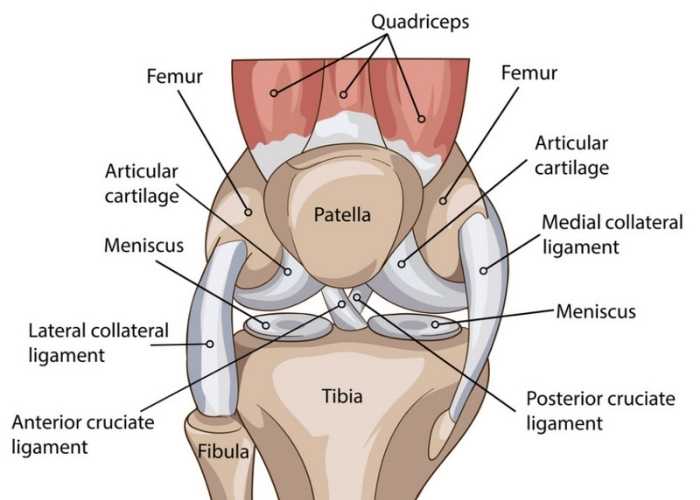
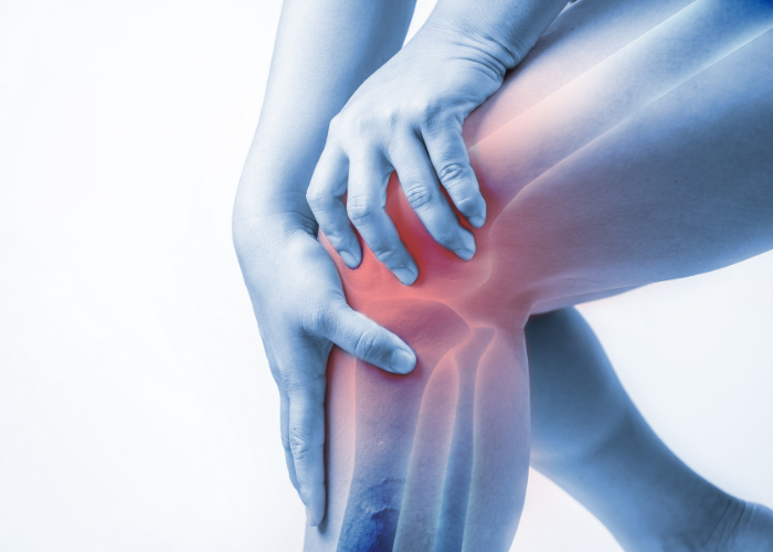
The connection of the femur (thigh bone), tibia (shin bone), and patella (kneecap) form the knee joint, one of the more complex human body joints. The patella is a sesamoid bone that is anchored in place by the quadriceps tendon attached to the femur and the patellar tendon attached to the tibia. A white, slippery connective tissue, known as articular cartilage, lines the undersurface of the patella allowing this bone to travel up and down the trochlear groove without pain. To make joint movement more seamless, the synovium is a thin layer of tissue that secretes a small amount of fluid to lubricate the articular cartilage. There is also a small fat pad that lies underneath the patella serving as a cushion and shock absorber. Chronic use of the knee joint with repetitive and intensive physical activities such as squatting and running can cause patellofemoral pain syndrome. This condition, also known as “Runner’s Knee”, occurs when the nerves of the knee joint perceive pain within the soft tissues surrounding the patella. Malalignment of the patella, quadriceps muscle weakness, and sudden changes to an exercise routine (i.e., intensity, frequency, or duration) can also lead to patellofemoral pain syndrome or anterior knee pain. Dr. Ronak Mukesh Patel, orthopedic knee specialist serving patients in Sugar Land, Pearland, and the Houston, Texas area, has the knowledge and understanding as well as substantial experience in treating patients who have experienced patellofemoral pain syndrome.
What are the symptoms of patellofemoral pain syndrome?
A common complaint of patellofemoral pain syndrome is knee pain that worsens with physical activity. Patients often describe a diffuse and dull pain in their knee that worsened gradually with time. Pain after sitting with the knees bent for a prolonged amount of time has also been reported. Some individuals also report a “crackling” or “popping” sound with knee movement after sitting for an extended period of time.
How is patellofemoral pain syndrome diagnosed?
Dr. Patel will obtain an extensive medical history, including physical activity habits, followed by a thorough physical examination of the knee joint. Patellofemoral pain syndrome is commonly diagnosed with a physical examination and medical history; however, diagnostic imaging tools such as x-rays and magnetic resonance imaging (MRI) may be requested to rule out damage to any other structures within the knee joint.
What is the treatment for patellofemoral pain syndrome?
Non-surgical treatment:
Non-operative therapies are often successful for most patients with a confirmed patellofemoral pain syndrome diagnosis. The pain and inflammation associated with this condition can be managed with a combination of RICE (rest, ice, compression, elevation) and non-steroidal anti-inflammatory medications (NSAIDs). Custom-made shoe insoles and taping the patella may be considered to help alleviate knee pain. Dr. Patel will recommend a physical rehabilitation program with a patellofemoral focus.
Surgical treatment:
Patellofemoral pain syndrome is rarely treated with surgical measures as the majority of patients are able to find relief with non-operative treatments. However, patients that continue to experience severe knee pain after a trial of conservative therapies may benefit from surgical intervention if structural problems are identified on MRI. A small camera (arthroscope) and specialized surgical instruments are used in a minimally invasive surgical procedure to identify the cause of knee pain. Dr. Patel will then implement one, or more, of the following procedures:
- The areas of tissue damage are excised and removed. Removal of any other abnormalities, such as loose bodies, inflamed tissues, or bone spurs, is also performed.
- Lateral Retinaculum Release. A fibrous tissue on the outer patella, known as the lateral retinaculum, is released as a common surgical technique for patellar instability. Releasing the lateral retinaculum can alleviate tension along the outer knee and help reposition the patella.
- Cartilage Transplant Surgery. If a cartilage defect is identified, this may be treated with cartilage restoration.
- Tibial Tubercle Osteotomy. This procedure is reserved for patients that experience patellar malalignment due to a shallow trochlear groove, patellar instability, or cartilage defects. This open surgical technique transfers the tibial tubercle (bony prominence of the shin bone), with the patellar tendon still attached, to another position on the tibia.
Patellar Tendon Pain Specialist

Do you have knee pain in and around your kneecap or patella? If so, you may be suffering from patellofemoral pain syndrome or patellar tendon pain. Patellofemoral pain syndrome specialist, Doctor Ronak Mukesh Patel, provides diagnosis as well as surgical and nonsurgical treatment options for patients in Houston, Sugar Land, and Pearland, TX who have suffered a patellar tendon tear, injury or have pain in their knee. Contact Dr. Patel’s team today!
