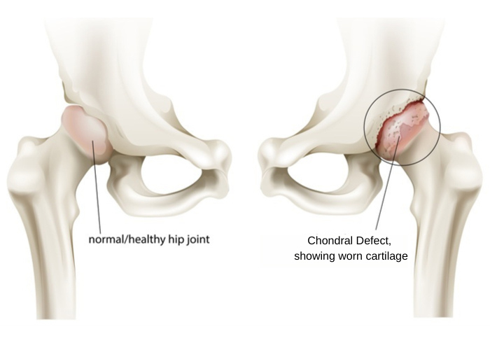
What is a chondral defect of the hip?
The articulation of the head of the femur (thigh bone) into the socket of the acetabulum (pelvis) forms the hip joint. The surface of the femoral head and acetabulum are blanketed in a shiny and slippery white tissue known as articular cartilage. This protective sheath of connective tissue allows the bones to painlessly glide over one another by reducing friction between these surfaces with hip movement. A chondral defect is a lesion of the articular cartilage that occurs with injury or damage. Due to the articular cartilage’s lack of blood supply, it has a very limited ability in repairing itself; therefore, degenerative joint diseases, such as arthritis, and joint pain can often develop.

What causes chondral defects of the hip?
The articular cartilage can deteriorate over time with natural wear-and-tear from age, activity level, and athletic activities. This natural tissue degeneration is frequently the cause of chondral defects in the hip. However, there are other mechanisms that can result in articular cartilage damage. Structural irregularities of the articulating bones, seen in femoroacetabular impingement (FAI), can produce chondral lesions. Blunt force trauma, such as a motor vehicle collision or a fall directly on the hip, can result in the articular cartilage breaking or tearing away from the bone. Athletes that repetitively use the hip joint in sports-related activities, such as soccer, football, hockey, and basketball, are vulnerable to experiencing chondral defects of the hip. Dr. Ronak Mukesh Patel, orthopedic hip specialist serving patients in Sugar Land, Pearland, and the Houston, Texas area, has the knowledge and understanding as well as substantial experience in treating patients who have experienced a chondral defect of the hip.
What are the symptoms of a chondral defect of the hip?
Individuals with a suspected chondral defect often report a dull, or intermittently severe, pain in the hip joint that worsens with physical activities. Some other symptoms of a chondral defect of the hip can include:
- Limping or abnormal gait can sometimes occur
- A substantial decrease in hip range of motion
- A “locking” or “catching” sensation with hip movement
How is a chondral defect of the hip diagnosed?
An interview will be conducted by Dr. Patel to gather a comprehensive medical history to include any prior hip injuries. A thorough physical examination will follow to evaluate the hip joint for pain, tenderness, and range of motion. X-rays may be requested if a hip injury resulted in chondral lesions. However, the best assessment tool to analyze the articular cartilage is magnetic resonance imaging (MRI).
What is the treatment for a chondral defect of the hip?
A number of factors, such as the patient’s age, activity level, medical history, size and number of lesions, and intended recovery goals, are taken into consideration by Dr. Patel and his orthopedic team when creating an individualized treatment plan.
Non-surgical treatment:
Patients with small chondral lesions may respond well to non-surgical therapies alone for symptom management. Limiting or modifying physical activities that worsen the hip pain is encouraged, although symptom relief is best achieved through avoiding these activities altogether. Pain and inflammation can also be managed by a combination of rest, ice, and non-steroidal anti-inflammatory medications (NSAIDs). A corticosteroid injection can be administered directly into the hip joint if pain persists with oral medications. Biologic treatments, such as the patient’s own platelet-rich plasma (PRP) or stem cells, can also be injected into the hip joint. When appropriate, a physical rehabilitation program may be prescribed to improve hip function and range of motion.
Surgical treatment:
Surgical intervention may be necessary for patients with severe articular cartilage damage or those that did not respond well to non-surgical therapies. Dr. Patel may implement one or more of the following surgical techniques to properly restore the articular cartilage:
- Chondroplasty: Also known as debridement, this arthroscopic technique uses specialized surgical instruments inserted through small incisions to excise and remove the chondral lesion(s).
- Microfracture: This method treats focal articular cartilage defects by drilling small openings into the bone. These openings allow stem cells and bone marrow to infiltrate the hip joint to stimulate new fibrocartilage growth over the exposed bone.
- Osteochondral Autograft/Allograft Transplantation Surgery (OATS): This surgical technique uses a cartilage graft from either the patient (autograft) or donor tissue (allograft) as a surface for new cartilage growth. The new tissue is molded to the patient’s specific chondral lesion(s) and situated as close to the native cartilage as possible.
Chondral Defects of the Hip Specialist

Are you an athlete or sports enthusiast who enjoys activities that require repetitive use of the hip joint, such as soccer, football, hockey, ballet, or basketball? If so, you may be at risk of developing a chondral defect of the hip. Chondral defects can cause pain, decrease in range of motion and early osteoarthritis. Hip preservation specialist, Doctor Ronak Mukesh Patel, provides diagnosis as well as surgical and nonsurgical treatment options for patients in Houston, Sugar Land, and Pearland, TX who are experiencing symptoms of a chondral defect. Contact Dr. Patel’s team today!


