An overview of the anatomy of the ankle:
The ankle is a complex hinge, synovial joint and consists of not only one joint, but two joints. The two joints are the subtalar joint and the tibiotalar joint. The difference in these joints is the movements they allow. The subtalar joint allows for side-to-side movement in the foot, while the true ankle joint allows up-and-down motion of the foot. Along with the bones in the ankle, there are also ligaments, muscles, and tendons. These components work together to handle the stress put on the ankle during walking, running, and jumping. The ankle is in constant motion, which makes it prone to injury. When the ankle suffers an injury due to overuse, trauma, or degeneration, patients may experience pain, loss of mobility, or ankle weakness. Dr. Ronak Mukesh Patel, orthopedic ankle specialist, treats patients in Sugar Land, Pearland, and the Houston, Texas area, who need an experienced doctor to diagnose and treat an ankle condition.
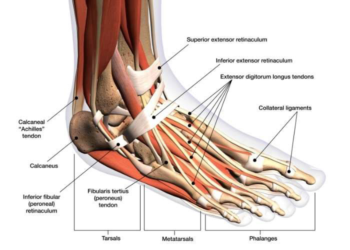
What is the anatomy of the ankle?
Bones of the ankle:
- Tibia: The shin bone or the medial (inside) portion of the ankle
- Fibula: The lower leg bone or lateral (outside) portion of the ankle
- Talus: The small bone sitting underneath the tibia and fibula
The lower ends of the tibia and fibula form a socket, where the talus sits. Without the talus, we wouldn’t be able to move our feet up and down.
Ligaments, muscles, and tendons of the ankle:
The ankle has many different ligaments, each with their own purpose and location:
- Deltoid Ligament: A thick ligament that supports the entire medial (inner) side of the ankle
- Distal Tibiofibular Syndesmosis
- Anterior Inferior Tibiofibular Ligament (AITFL): Connects the tibia to the fibula in the back of the tibia and fibula
- Posterior Inferior Tibiofibular Ligaments (PITFL): ligaments which crisscross the back of the tibia and fibula
- Inferior transverse ligament
- Interosseous Ligament & Membrane: Rests between the tibia and fibula and runs the entire length of the tibia and fibula, from the knee to the ankle
- Lateral Ligament complex: Attached to the lateral malleolus (a bony prominence located on the lateral end of the fibula). The lateral ligament is responsible for resisting over-inversion (turning inward) of the foot. The lateral ligament complex consists of three ligaments:
- Anterior talofibular ligament (ATFL): Connects the lateral malleolus to the lateral end of the talus
- Posterior talofibular ligament (PTFL): Connects the lateral malleolus and the posterior end of the talus
- Calcaneofibular ligament (CFL): Connects the lateral malleolus and the calcaneus (heel)
Muscles and tendons:
- Peroneal tendons: Responsible for allowing the ankle to bend inward and outward
- Posterior Tibialis tendon: Allows the ankle to turn inward, while also supporting the arch of the foot
- Anterior Tibialis tendon: Allows the ankle and foot to turn upward
- Achilles tendon: Connects the calf muscles to the calcaneus (heel)
Ankle Cartilage:
Articular cartilage covers the ends of each bone in the ankle and allows the bones to move smoothly against each other. The cartilage helps the joint carry weight by providing shock absorption.
Dr. Patel treats these common Ankle Conditions:
Ankle Expert

The foot and ankle is complex hinge, synovial joint and consists of 2 joints. Various bones, ligaments, tendons and muscles give the ankle power, motion and stability. When the ankle or foot suffers an injury, it is important to see an ankle expert who has extensive experience in treating ankle conditions and injuries. Ankle expert, Doctor Ronak Mukesh Patel, provides diagnosis as well as surgical and nonsurgical treatment options for patients in Houston, Sugar Land, and Pearland, TX who have sustained a foot or ankle injury. Contact Dr. Patel’s team today!