What is the hip joint?
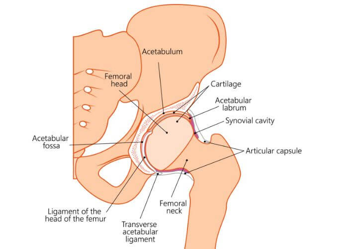
The hip joint connects the lower limbs to the trunk of the body and is the largest weight-bearing joint within the human body. This joint has a ball-and-socket joint structure that is formed by the head of the femur (thigh bone) joining the socket of the acetabulum (pelvis). The hip joint has large muscles, tendons, and ligaments that are connected to the femur and pelvic bone to prevent joint dislocation. Because of the stress continually encountered by the hip joint, any injury or disease can alter its ability to function properly. When hip conditions are left untreated, they can often become debilitating resulting in difficulties performing activities of daily living or enjoying sports-related activities. Dr. Ronak Mukesh Patel, orthopedic hip specialist serving patients in Sugar Land, Pearland, and the Houston, Texas area, has the knowledge and understanding of hip anatomy as well as substantial training and experience in treating common hip injuries.
What are the important structures of the hip joint?
The hip joint is an intricate arrangement of bones, muscles, ligaments, nerves, and arteries. A healthy hip joint is vital for participating in athletic activities as well as performing daily activities such as running and walking.
Bones
Femur: This bone is found in the thigh and is the longest of the 206 bones within the body. The femoral head joins the acetabulum socket of the pelvis to create the hip joint. Near the neck of the femur are two bony prominences, the greater and lesser trochanters, that serve as muscle attachment sites.
Pelvis: There are three bones that form the pelvis: the ilium, the ischium, and the pubis. The acetabulum is a deep circular socket on the outer edge of the pelvis that is formed from the union of these three bones. This socket is joined with the head of the femur to form the hip joint. The acetabulum of the pelvis and surrounding muscles and ligaments work collectively to maintain the stability of the hip joint.
Cartilage
Articular Cartilage: Surrounding the head of the femur and coating the surface of the acetabulum socket is a shiny and slippery white tissue known as articular cartilage. This connective tissue reduces friction with joint movement and also provides a protective barrier for these bones.
Acetabular Labrum: Lining the socket of the acetabulum is a ring of cartilage tissue that further deepens the acetabular cavity in addition to enhancing the stability and strength of the hip joint.
Synovial Membrane: There is a thin joint capsule found within the joint space that secretes a thick viscous fluid to lubricate the hip joint and reduce friction with hip joint movement.
Bursae: These small, fluid-filled sacs are important for preventing inflammation of the major structures of the hip joint by reducing friction between the tissues with hip joint movement.
Ligaments
Similar to strong ropes, the ligaments of the hip joint are elastic bands of tissue that connect two bones and maintain the alignment of these bones. These ligaments form a dense and fibrous network of connective tissues surrounding the joint capsule.
Ischiofemoral Ligament: This triangular-shaped ligament band limits internal rotation of the hip. It originates from the ischium and the posterior acetabulum and combines with the fibers of the joint capsule.
Pubofemoral Ligament: This ligament prevents hyper-abduction of the hip joint. Its triangular shape can be found on the underside of the hip joint connecting the pubis to the femoral head.
Iliofemoral Ligament: This Y-shaped ligament is the strongest one in the body and limits over-extension of the hip. It connects the pelvis and femoral head and is located at the front of the hip joint.
Ligamentum Teres Femoris: Between the acetabulum and femoral head is where this flattened ligament can be found. Although it does not contribute to hip movement, it does house the arterial blood supply for the head of the femur.
Muscles
Even though the muscles of the hip can be divided into five groups, they share a similar function of stabilizing the pelvis by acting on the thigh.
Iliopsoas: This muscle group contains the iliacus, psoas major, and psoas minor muscles. They originate from the posterior abdominal wall and attach to the lesser trochanter on the femur. This muscle group has a role in flexion and external rotation of the thigh.
Gluteals: There are superficial and deep gluteal muscles. The gluteus maximus, gluteus medius, gluteus minimus, and tensor fascia lata make up the superficial muscle group which enables extension and thigh movement away from the midline. The deep gluteal muscle group consists of the quadratus femoris, gemellus inferior, gemellus superior, piriformis, obturator internus, and obturator externus. This muscle group stabilizes the pelvis with external rotation of the thigh at the hip joint.
Adductors: There are six hip adductor muscles that form this muscle group: the pectineus, gracilis, adductor longus, adductor brevis, adductor magnus, and adductor minimus. These muscles originate from the pubis, travel over the hip joint, and attach to the femur. The adductor muscles facilitate thigh movement towards the midline of the body.
Rectus Femoris: This is one of the four quadriceps femoris muscles located on the anterior (front) thigh. It is a hip flexor muscle that originates at the ilium of the pelvis. This muscle flexes the thigh at the hip joint as well as leg extension at the knee joint.
Hamstrings: The semitendinosus, semimembranosus, and biceps femoris are the three biarticular muscles that form the hamstrings. These muscles originate at the bottom of the pelvis and travel down the posterior (back) thigh. This muscle group pulls the hip backward to aid in hip extension and also contributes to knee flexion.
Nerves
There are three nerves associated with the hip: the femoral nerve, the sciatic nerve, and the obturator nerve. These nerves receive instructions for muscle movement through signals from the brain. They are also responsible for sending sensory information, such as pain, touch, and temperature, back to the brain.
Arteries
The lower limbs are saturated with a healthy supply of blood vessels. The main arterial supply for the lower limb is the femoral artery, which emanates from the deep pelvis into the anterior thigh. The femoral artery is one of the largest in the body and can be palpated through the skin of the upper thigh.
What causes hip pain?
Hip pain is associated with several different conditions. Osteoarthritis and rheumatoid arthritis are the most common among older adults. Hip pain is a frequently reported symptom in the following conditions:
How is hip pain treated?
Dr. Patel and his orthopedic team will review a number of factors, such as the patient’s age, activity level, medical history, injury severity, and desired recovery goals, when designing an appropriate treatment plan.
Non-surgical therapies alone often work well for treating hip pain. These treatment measures can include activity modification, ice, non-steroidal anti-inflammatory medications (NSAIDs), and a physical therapy program. However, surgical treatment may be necessary when non-surgical therapy fails or a patient sustains a severe hip injury. A minimally invasive arthroscopic procedure can be performed with a small camera (arthroscope) and specialized surgical instruments to conduct the necessary revisions. This surgical technique is often favored by surgeons and patients alike as it reduces the risk of infection and blood loss during the surgery and provides the best recovery outcomes.
Hip Expert

The hip is an intricate joint that is the largest weight-bearing joint in the human body. This joint is composed of a number of muscles, tendons, ligaments, and bones. Injuries to any part of this joint must be treated appropriately in order to restore and maintain a normal range of motion and stability. Complex hip expert Dr. Ronak Mukesh Patel is extremely knowledgeable in diagnosing and treating patients in Houston, Sugar Land, and Pearland, TX who have experienced a hip injury or condition. Contact Dr. Patel’s team today!