What is a lateral collateral ligament injury?
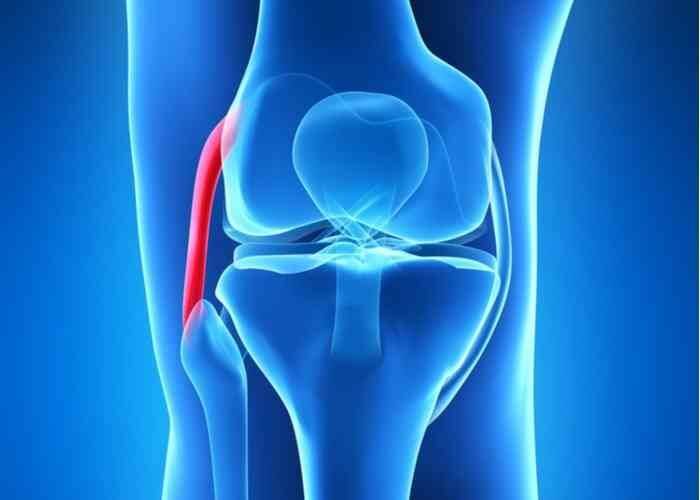
There are two collateral ligaments within the knee complex that are located outside of the knee joint (extra-articular). The lateral collateral ligament (LCL) is found along the outer edge of the knee and emanates from the lateral epicondyle, a bony prominence on the outer femur (thigh bone) and attaches to the head of the fibula (smaller bone alongside the tibia). Because the LCL is anchored on the fibular head, it is also known as the fibular collateral ligament (FCL). The LCL works collectively with the medial collateral ligament (MCL) to protect the knee joint against side to side movements and provide rotational stability. A considerable force placed on the inner knee can cause the knee joint to shift sideways resulting in an LCL injury. These ligament injuries are graded according to the amount of damage sustained and can range from stretching of the LCL to a complete separation from its attachment site. Injuries to the LCL are also frequently associated with multiple ligament injuries including the anterior cruciate ligament (ACL), posterior cruciate ligament, MCL, and posterolateral corner (PLC). These injuries are also associated with nerve injuries involving the peroneal nerve and rarely a blood vessel (vascular) injury that can be serious. It is crucial to have this closely evaluated. Dr. Ronak Mukesh Patel, orthopedic knee specialist serving patients in Sugar Land, Pearland, and the Houston, Texas area, has the knowledge and understanding, as well as substantial experience, in treating patients who have experienced a lateral collateral ligament injury.
Are certain individuals at a higher risk of a lateral collateral ligament injury?
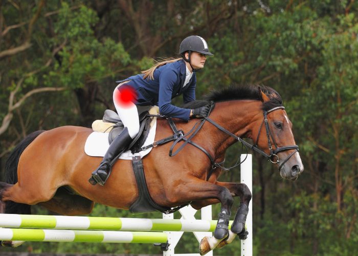
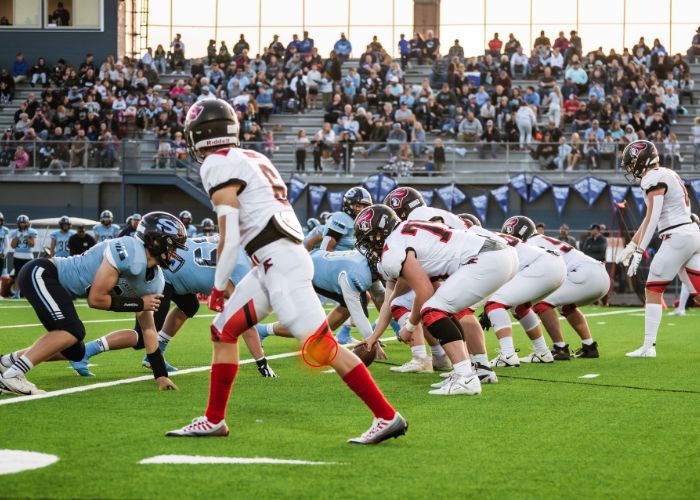
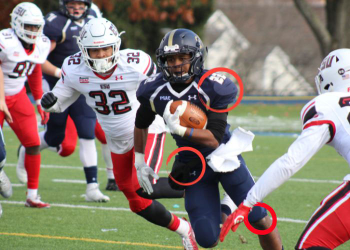
Lateral collateral ligament (LCL) injuries frequently occur from sports-related activities, therefore athletes are the most susceptible to developing an LCL injury. Some examples that can result in an LCL injury are side tackles in football, jumping and rapid direction changes seen in basketball, and the frequent stop-and-go and weaving motions with soccer. The recurrent bending and twisting motions associated with gymnastics, cheerleading, and skiing can lead to an LCL injury.
What are the symptoms of a lateral collateral ligament injury?
Patients with a suspected lateral collateral ligament (LCL) injury frequently report pain and tenderness along the outer edge of the knee immediately following an injury. This outer knee pain may also worsen with time. Some other common symptoms of an LCL injury include:
– Decreased range of motion
– Visible bruising and swelling along the outer edge of the affected knee
– Knee joint Instability, especially with more severe tears
– A “locking” or “buckling” sensation with knee joint movement
How is a lateral collateral ligament injury diagnosed?
A comprehensive medical history is gathered through an interview with Dr. Patel and will include information pertaining to the precipitating injury, any prior knee injuries, or any other underlying health conditions. This interview is followed by a thorough physical examination where Dr. Patel will evaluate range of motion of the knee and assess for any knee instability. Diagnostic imaging studies are often beneficial in confirming a lateral collateral ligament (LCL) injury diagnosis. While x-rays can identify any bone-related injuries, magnetic resonance imaging (MRI) is the best diagnostic imaging tool for LCL injury confirmation.
What is the treatment for a lateral collateral ligament injury?
Non-surgical treatment:
Patients with a confirmed mild lateral collateral ligament (LCL) injury often benefit from conservative therapies alone. A knee brace is important for protecting the knee joint from further damage while allowing the ligaments to properly heal. The use of crutches or a walker limits weight-bearing onto the affected knee. The pain and inflammation associated with an LCL injury can be managed with a combination of RICE (rest, ice, compression, elevation) and non-steroidal anti-inflammatory medications (NSAIDs). Dr. Patel will provide instructions for stretching and range of motion exercises to perform at home when appropriate.
Surgical treatment:
Patients with more severe lateral collateral ligament (LCL) injuries or those that did not benefit from conservative therapies may require surgical intervention to restore stability to the knee joint. Surgical repair of the LCL necessitates surgical repair or reconstruction of the entire ligament. For a repair, the damaged ligament fragments are resected, and the remaining healthy tissue is sutured back together or securely anchored to the bone. For reconstruction, a tissue graft is harvested, either from the patient (autograft) or donor (allograft) and sewn into the native tissue before reattaching the ligament back in the correct anatomical position. This surgery is done in combination with a minimally invasive arthroscopic procedure utilizing a small camera (arthroscope) and specialized surgical instruments. Often times, associated ACL, PCL, MCL, and/or PLC injuries are also addressed with this surgery.
LCL Injury Specialist

The LCL is a ligament in the knee that protects the joint during side-to-side and twisting movements. Individuals that participate in activities involving these motions such as soccer, football, or tennis are at an increased risk of injuring the LCL. Depending on the severity of the tear or damage, treatment may require surgery. Complex knee specialist, Doctor Ronak Mukesh Patel, provides diagnosis and surgical or nonsurgical treatment options for patients in Houston, Sugar Land, and Pearland, TX who have suffered an LCL injury. Contact Dr. Patel’s team today!
