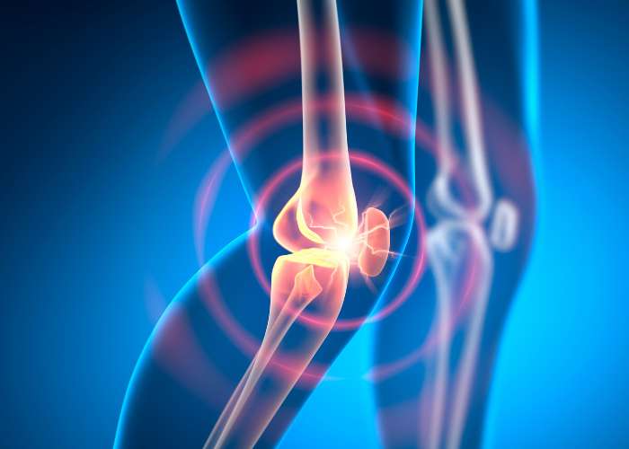
What is patellar instability?
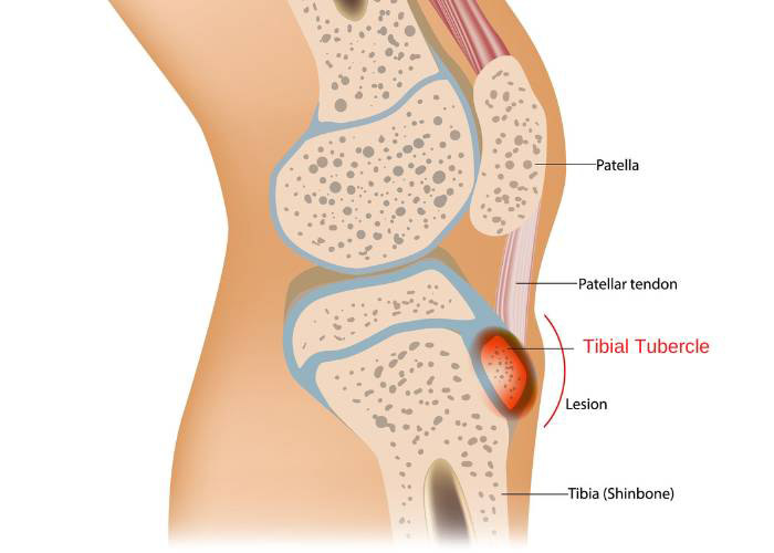
The knee joint is formed by the connection of the lower femur (thigh bone), upper tibia (shin bone), and patella (kneecap). The patella is a sesamoid bone that is anchored within the anterior knee by the quadriceps tendon extending from the femur and the patellar tendon from the tibia. A traumatic event, such as a direct and forceful blow to the kneecap or abnormal twisting of the knee, can cause the patella to be dislodged from its normal position. This injury, known as a patella dislocation, can also occur from underlying knee abnormalities such as an elevated kneecap or muscle weaknesses. The tendons anchoring the patella in place can be stretched or torn from patellar displacement. Any damage sustained by these tendons can lead to ligament laxity further increasing the likelihood of future recurrent patellar dislocations.
What is patellar malalignment?
The patella moves along the trochlear groove, a bony channel on the distal femur, as the knee bends and straightens. This bony channel can occasionally be more shallow in some individuals and result in displacement of the patella from a minimal force. The patellar tendon is attached to the tibial tubercle, a bony prominence on the shin bone. Patellar malalignment also occurs when this tibial tubercle is not positioned correctly with a shallow trochlear groove.
What is the treatment for patellar instability or patellar malalignment?
Patients with a patella dislocation or a history of recurrent patella dislocations do not typically benefit from non-operative therapies alone. The best outcome for re-establishing patellar stability and/or correcting patellar malalignment occurs with a tibial tubercle osteotomy. This procedure repositions the tibial tubercle on the shin bone to ensure proper tracking of the kneecap by pulling the patella inward. Dr. Ronak Mukesh Patel, orthopedic knee doctor, treats patients in Sugar Land, Pearland, and the Houston, Texas area, who have experienced patellar instability or patellar malalignment and are in need of a tibial tubercle osteotomy.
How is a tibial tubercle osteotomy performed?
A tibial tubercle osteotomy involves two surgical techniques that are performed in the same setting. The arthroscopy technique involves a small camera (arthroscope) and specialized surgical instruments to make the necessary corrections. Dr. Patel uses the arthroscope to methodically examine the cartilage, menisci, and ligaments of the knee joint. The surgical instruments are then inserted to address any conditions documented during the examination.
When the necessary repairs, if any, are completed, a four to six-inch incision is created over the proximal tibia to begin the osteotomy portion of the surgery. A surgical saw or bone chisel is introduced to detach the tibial tubercle from the tibia. Once the tibial tubercle is separated with the patellar tendon still connected, the resected bone is typically repositioned and anchored to the tibia with metal screws. The devices used in securing the tibial tubercle are permanent; however, these can be removed at a later date if they generate any pain or discomfort. To ensure proper bone alignment, the tracking pattern of the patella is confirmed.
What is the recovery period like after a tibial tubercle osteotomy?
The recovery period following a tibial tubercle osteotomy is often a longer and more involved process. Most patients can expect a return to non-impact activities in 3 to 4 months with a full recovery to normal physical activities in approximately 6 to 8 months. Compliance with the post-operative care instructions set forth by Dr. Patel can ease the recovery and improve the probability of a more successful outcome. Patients in the Houston, Texas area can typically anticipate the following during the recovery period:
- The knee is immobilized by a cast or other device immediately following surgery. Weight-bearing may be limited for about 6 weeks.
- The stress loaded onto the repaired knee should be limited until the osteotomy site has completely healed. There is a greater risk of fracturing the tibia if the patient places a significant amount of stress before the osteotomy has completely healed.
- Adhering to and completing the physical rehabilitation program designated by Dr. Patel to strengthen and restabilize the knee joint. Passive exercises begin after surgery and gradually progress as appropriate.
Tibial Tubercle Osteotomy Surgeon

A tibial tubercle osteotomy can be helpful if the patella (kneecap) easily dislocates, or if there is knee malalignment. This type of osteotomy can restore the patella’s stability int he knee. Tibial tubercle osteotomy surgeon, Doctor Ronak Mukesh Patel, provides diagnosis as well as surgical and nonsurgical treatment options for patients in Houston, Sugar Land, and Pearland, TX who have knee malalignment or a patella that dislocates easily. Contact Dr. Patel’s team today!