FAI Hip Impingement Specialist

Femoroacetabular impingement is a hip condition common in active individuals and people diagnosed with degenerative joint disease. This condition causes bone spurs to develop in the hip socket which rub and cause discomfort with movement. Hip specialist Dr. Ronak Mukesh Patel provides diagnosis and specialized treatment plans for patients in Houston, Sugar Land, and Pearland, TX. Contact Dr. Patel’s team today!
What is FAI hip impingement or femoroacetabular impingement?
The head of the femur (thigh bone) connects with the socket of the acetabulum (pelvis) to form the ball-and-socket type of hip joint. A white slippery sheath of articular cartilage protects the ends of these bones and enables joint movement with very little friction. This cartilaginous covering can be subject to premature erosion when the bones are not aligned properly. In femoroacetabular impingement (FAI), the femoral head and/or the acetabular socket develop bony overgrowths, called bone spurs, that rub against the articular cartilage preventing seamless joint movement.
These bone spurs gradually weaken the articular cartilage leading to osteoarthritis and can also tear the hip labrum surrounding the acetabular socket. FAI hip impingement can begin as early as childhood and is the leading factor of early-onset degenerative joint disease in young and active individuals. Dr. Ronak Mukesh Patel, orthopedic hip pain specialist serving patients in Sugar Land, Pearland, and the Houston, Texas area, has the knowledge and understanding as well as substantial experience in treating patients who have experienced femoroacetabular impingement.
Are there different forms of FAI hip impingement?
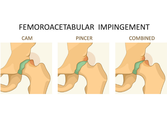
Individuals who experience femoroacetabular impingement (hip FAI) may be diagnosed with one of two conditions or a combination of the two.
- Cam Impingement: When the femoral head is irregularly shaped then it is unable to rotate properly inside the acetabular socket. Patients with cam impingement have a structural abnormality of this femoral head which leads to excessive rubbing against the articular cartilage and can result in a hip labrum tear. This is also known as a pistol grip deformity.
- Pincer Impingement: Patients with this condition have an extra amount of bone that extends past the standard rim of the acetabulum. Normal hip flexion can cause the femoral neck to knock against this bony protrusion and lead to a crushed hip labrum or articular cartilage damage. This is more commonly seen in females.
- Combined Impingement: This condition is diagnosed when both the cam and pincer types of impingements are present. This is the most common form of impingement.

What are FAI hip symptoms?
A common complaint of FAI hip impingement is a constant and sharp pain in the groin (front) area of the hip that worsens with internal rotation and flexion of the hip. However, it is important to note that individuals may not report any symptoms until there is damage to the hip labrum and/or articular cartilage. Additional symptoms of hip FAI can include:
- Hip stiffness
- Decreased range of motion
- Groin pain or pain in the front of the hip
- Pain that radiates to the outer hip
- Abnormal gait
- Pain that fluctuates between a dull ache and intense stabbing
- Pain with sitting for long periods
- Pain with squatting
- Pain with sports
How is hip FAI diagnosed?
Dr. Patel will gather a complete medical history followed by a thorough physical examination. During the exam, Dr. Patel will bring the knee up to the chest and rotate it towards the opposing shoulder in a maneuver known as the impingement test or FADIR (flexion, adduction and internal rotation) test. A positive hip impingement test is indicated by the re-creation of hip pain with hip rotation. Another positive indicator of FAI is if the hip pain resolves after injection of a local anesthetic (numbing medicine) with or without steroid into the hip joint. This is done in Dr. Patel’s office under ultrasound guidance or during the same setting as an MRI. Diagnostic imaging studies are also beneficial for diagnosing FAI. X-rays can determine if any bony abnormalities are present including impingement and arthritis with bone spurs. Magnetic resonance imaging (MRI) can identify the extent of damage to the hip labrum and/or articular cartilage as well as evaluate the surrounding soft-tissue structures for additional injury. A computed tomography (CT) scan is often utilized to visual the bony structure more in-depth in 3D utilizing the Stryker HipMap technology. This allows in-depth analysis of the hip morphology including femoral and acetabular version.
What is FAI treatment for hip impingement?
Non-surgical treatment:
This is the initial treatment of choice in all patients. Patients that experience mild femoroacetabular impingement (FAI) symptoms may find relief with non-surgical treatment measures. Avoiding activities that exacerbate pain is recommended. A combination of non-steroidal anti-inflammatory medications (NSAIDs) with periodic hip injections can provide long-term pain and inflammation relief. Physical therapy with specific hip/core mobility and strengthening exercises can mitigate the stress on the injured cartilage or labrum.
Surgical treatment:
When non-surgical treatment measures fail to alleviate the symptoms of FAI hip impingement, surgical intervention may be needed. Hip preservation surgery is done with minimally invasive (arthroscopic) technique. A postless technique is utilized with a special operating room table. Dr. Patel performs surgeries at Texas Orthopedic Hospital in Houston, TX which is a world-class, specialty orthopedic hospital in the heart of the Texas Medical Center. = Femoroplasty or osteochondroplasty (for cam impingement) and acetabuloplasty (for pincer impingement) are performed with minimally invasive arthroscopic technique, that use a small camera (arthroscope) and specialized surgical instruments, and are preferred by patients and surgeons alike. When the injured labrum is repaired and damaged articular cartilage fragments are excised and removed. Depending on the specific type of FAI, the femoral head is reshaped, or the rim of the acetabular socket is trimmed to the normal shape. Surgical treatment of FAI is important for hip preservation to slow down further damage to the articular cartilage and/or hip labrum and return to normal activity.
Link


