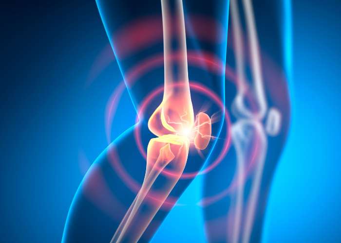
What is the knee joint?
Of the 360 joints within the human body, the knee is the largest and most complex joint. The knee’s anatomically intricate design enables it to sustain large amounts of internal and external stress every day. The knee can flex, extend, and twist side-to-side; however, this mobility can make the knee joint vulnerable to injury. Dr. Ronak Mukesh Patel, orthopedic knee specialist serving patients in Sugar Land, Pearland, and the Houston, Texas area, has the knowledge and understanding of knee anatomy, as well as substantial training and experience in treating common knee conditions and injuries.

What are the important structures of the knee joint?
Bones, cartilage, ligaments, and tendons are the four main elements of the knee joint. Other tissues, such as the muscles and bursae, are vital for providing mobility and strength to the knee joint.
Bones:
The knee joint is formed by the articulation of the lower end of the femur (thigh bone), the upper end of the tibia (shin bone), and the patella (kneecap). A smaller bone (fibula) alongside the tibia holds an attachment site for an extra-articular ligament. The long bones (tibia and femur) assist in joint movement and bearing weight, whereas the patella, which sits in front of the femur and tibia, protects the intra-articular ligaments and helps straighten the knee joint.
Cartilage:
There are two main types of cartilage in the knee joint:
- Articular Cartilage: The ends of the femur and tibia as well as the posterior (back) patella are blanketed in a shiny and slippery white tissue known as articular cartilage. This connective tissue gives these bones a layer of protection while enabling the ends of these bones to painlessly slide over one another as the knee bends and straightens.
- Meniscus: There are two crescent-shaped pieces of meniscus cartilage found between the femur and tibia. This tough and rubbery tissue differs from articular cartilage as it reduces friction between the femur and tibia by acting as a shock absorber. The medial meniscus, found along the inner knee, sustains up to 50% of the stress exerted on the inside of the knee. The lateral meniscus, found along the outer knee, sustains up to 80% of the stress exerted on the outside of the knee. The menisci evenly distribute this weight between the femur and tibia to provide stability and protection of the soft-tissue structures of the knee joint. The meniscus cartilage can be divided into two portions: the “red zone” and “white zone”. The outer third of the meniscus, the “red zone”, contains a healthy blood supply allowing injuries and damage to potentially heal. The inner two-thirds, the “white zone”, lacks this blood supply necessary for reliable tissue repair. The meniscus cartilage also protects the articular cartilage to prevent early and accelerated tissue degeneration causing early-onset arthritis.
Ligaments:
Within the knee joint are elastic bands of tissue (ligaments) that are responsible for connecting bones and maintaining bone alignment. These ligaments also stabilize the knee joint. There are four main ligaments within the knee joint that are classified as cruciate and collateral ligaments.
- Cruciate Ligaments: The cruciate ligaments found within the knee joint are responsible for limiting forward and backward movement of the tibia in relation to the femur. The anterior cruciate ligament (ACL), frequently damaged with twisting movements, travels from the posterior (back) femur through the knee joint and attaches to the anterior (front) tibia. In conjunction with the ACL, the posterior cruciate ligament (PCL) forms an “X” by traveling from the anterior femur to the posterior tibia. Of the four main knee ligaments, the PCL is the strongest and is injured less frequently. A PCL injury is typically a secondary injury due to the amount of force required to perpetrate ligament damage.
- Collateral Ligaments: The collateral ligaments are located outside of the knee joint (extra-articular) and are responsible for protecting against unusual twisting motions and stabilizing the knee. The medial collateral ligament (MCL) prevents the knee from collapsing inward and originates along the inner femur and attaches to the inner tibia. The lateral collateral ligament (LCL), or fibular collateral ligament (FCL), prevents the knee from collapsing outward and originates from the lateral epicondyle, a bony prominence on the outer femur, and attaches to the head of the fibula.
Tendons:
There are tough bands of tissue (tendons) that connect muscle to bone and stabilize the knee joint. The largest tendon of the knee joint is the quadriceps tendon that connects the quadriceps femoris muscle group of the anterior (front) thigh to the patella. The patellar tendon connects the patella to the tibia thus acts similarly to a ligament. Working together, the quadriceps tendon and the patellar tendon straighten and bend the knee.
Bursae:
There are small, fluid-filled sacs within the knee joint that secrete a thick viscous fluid to further reduce friction between the bones and tissues with joint movement. These sacs, known as the bursae, also help prevent inflammation of the major structures of the knee joint. There are approximately fourteen bursae in each knee joint.
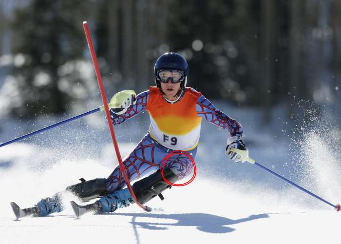
What are common knee injuries?
The knee joint is at risk for injury because of the multiple structures that are intricately connected. Damage to the knee can be inflicted by sports-related activities, repetitive use, a degenerative condition, or even a simple accident such as a fall. The knee injuries commonly treated by Dr. Patel are, as follows:
- Cartilage Injuries of the Knee
- Meniscus Tears of the Knee
- Patellar Tendon Injury
- Patella Dislocation
- Anterior Crucial Ligament (ACL) Injury
- Posterior Cruciate Ligament (PCL) Injury
- Medial Collateral Ligament (MCL) Injury
- Medial Patellofemoral Ligament (MPFL) Injury
How are knee injuries treated?
There are a number of common knee injuries that can be treated with non-surgical therapies alone. However, these treatment measures might be unsuccessful for more severe knee joint injuries thus requiring surgical intervention. Dr. Patel and his orthopedic team will confirm if surgery is needed and identify the appropriate surgical approach to best treat the patient’s specific injury. Dr. Patel favors the minimally invasive arthroscopic approach when surgically repairing a knee injury. This surgical technique involves a small camera (arthroscope) and specialized surgical instruments to conduct the necessary revisions and has been shown to as an effective treatment measure for providing the best recovery outcomes.
Knee Expert

The knee is the largest and most complex joint in the human body. The knee has the ability to flex, extend, and twist side to side which makes it incredibly flexible. However, the flexibility of the knee makes it vulnerable to injury. Some of the most common injuries of the knee include arthritis, ACL tears, MCL tears, and meniscus or knee cartilage tears. Knee specialist, Doctor Ronak Mukesh Patel, provides diagnosis as well as surgical and nonsurgical treatment options for patients in Houston, Sugar Land, and Pearland, TX who have sustained a knee injury. Contact Dr. Patel’s team today!